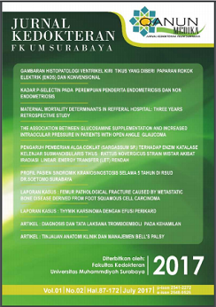
Histopathology of Left Ventricular Mice Presented with Electrical Cigarettes (ENDS) and Conventional
Abstract
ABSTRACT
Electric Cigarette (ENDS) and conventional contains carbon monoxide (CO) which is a major cause of cardiovascular disease. CO is a dangerous substance since as free radicals it have high affinity for hemoglobin. Thus affinity could lead to tissue hypoxia resulting in cell death (necrosis) started by the cell nucleus pyknosis. In order to further study about the effect, this experiment is conducted to identify the histopathological appearence  of the left ventricle rat against exposure to electric cigarette (ENDS) and conventional. This study was conducted with a post test only control group design method. Samples contained 18 Rattus novergicus rat which was divided in 3 groups: control group (K) as a negative control, P1 group were exposed by conventional cigarretes, and P2 group were exposed by ENDS. The treatment had been maintained for 30 days, followed by termination to perform the heart organ dissection and HE staining in order to made preparat. Preparat was observed by 400x lens of microscope to identify the pyknosis of the nuclei in 5 field of view. Data were analyzed with oneway ANOVA test with p<0,05 and CI=95%. This study proved that there was a significant differences of pycnotic nuclei amount among the group (p-value=0,008). There was significant difference between group K with P1 (p=0,004), group K with group P2 (p=0,012). There was no significant difference between group P1 and P2 (p=0,567).It can be conluded that exposure to either electric cigarette (ENDS) or conventional cigarette gives significant effect to cell nucleus pycnosis. Meanwhile, the effect comes from exposure of electric cigarette smoke (ENDS) has no significant difference with conventional cigarette.ÂKeywords: histopathological appearance of left ventricle, electrical cigarette, ENDS, conventional cigarette
Â
ABSTRAK
Rokok elektrik (ENDS) dan konvensional mengandung karbonmonoksida (CO) yang diduga berperan utama menyebabkan penyakit kardiovaskuler.CO merupakan zat yang berbahaya karena sebagai radikal bebas dan sifatnya yang memiliki afinitas yang tinggi terhadap Hemoglobin. Hal ini dapat menyebabkan hipoksia jaringan yang mengakibatkan kematian sel (nekrosis) ditandai dengan inti sel yang piknosis. Tujuan penelitian ini untuk mengidentifikasi gambaran histopatologi ventrikel kiri tikus terhadap paparan rokok elektrik (ENDS) dan konvensional. Penelitian ini merupakan penelitian eksperimental dengan rancangan post test only control group design. Sampel adalah tikus Rattus norvegicus dengan jumlah 18 ekor dibagi dalam 3 kelompok, yakni kelompok K sebagai kontrol negatif, kelompok P1 diberikan paparan Rokok Konvensional dan kelompok P2 diberikan paparan rokok elektrik (ENDS). Sampel diperlakukan selama 30 hari, kemudian diterminasi untuk pengambilan organ jantung dan dibuat preparat dengan pengecatan HE. Pembacaan preparat dengan mikroskop perbesaran 400x untuk melihat inti sel piknotik pada 5 lapang pandang. Data dianalisis menggunakan uji Oneway ANOVA dengan p<0,05 dan CI=95%. Dari hasil penelitian didapatkan perbedaan yang bermakna jumlah inti sel piknotik pada ketiga kelompok (p-value=0,008). Terdapat perbedaan yang signifikan antara kelompok K dan P1 (p=0,004), kelompok K dan P2 (p=0,012). Namun antar kelompok P1 dan P2 tidak terdapat perbedaan bermakna (p=0,567).Paparan rokok elektrik dan konvensional memiliki efek yang signifikan dibanding kontrol terhadap jumlah inti piknotik. Namun, Jumlah inti sel piknotik ventrikel kiri tikus tidak mengalami perbedaan bermakna antara paparan asap rokok elektrik (ENDS) dan konvensional.
Â
Kata kunci: Histopatologi ventrikel kiri, rokok elektrik,ENDS, rokok konvensional.
Â
Â
Full text article
References
Ambrose JA, Rajat SB. The Pathophysiology of Cigarette Smoking and Cardiovaskular Disease. J Am Coll Cardiol. 2004;43(10):1731-7.
Anggraeni, N.I. S. Pengaruh Lama Paparan Asap Knalpot Dengan Kadar CO 1800 PPM Terhadap Gambaran Histopatologi Jantung Pada Tikus Wistar. [Tesis]. 2009. Semarang : Fakultas Kedokteran Univesitas Diponegoro.
Armani C., Landini L., Jr, Leone A. Molecular and biochemical changes of the cardiovascular system due to smoking exposure. Curr. Pharm. Des. 2009;15:1038–1053. doi: 10.2174/138161209787846973.
Bernhard D., Wang X.L. Smoking, oxidative stress and cardiovascular diseases—Do anti-oxidative therapies fail? Curr. Med. Chem. 2007;14:1703–1712. doi: 10.2174/092986707781058959.
Dharmawan, Teddy. Putra, Uji Toksisitas Akut Monocrotophos Dosis Bertingkat Per Oral Dilihat Dari Gambaran Histopatologis Otot Jantung Mencit BALB/C. [Skripsi]. 2010. Semarang : Fakultas Kedokteran Universitas Diponegoro;.
El Morsy A, Amel As, Manal Hm. Histologicaland Scanning Electron Microscopic Study Of The Effect Of Cigarette Smoke On The Heart Of Male Albino Rats. The Egypt J of Histo. 2007;30 (2):409-18.
Farsalinos KE, Giorgio R,Elena A,Emiliano R,Elena B,Stefano T et al. Comparison of the Cytotoxic Potential of Cigarette Smoke and Electronic Cigarette Vapour Extract on Cultured Myocardial Cells. Int J Environ Res Public Health. 2013; 10(10): 5146–5162. doi: 10.3390/ijerph10105146
Fitria,Retno T, Jubhar CM, Ferry FK. Merokok dan Oksidasi. Sains Medika.2013;5(2):113-20.
Izzotti A., D’Agostini F., Balansky R., Degan P., Pennisi T.M., Steele V.E., De Flora S. Exposure of mice to cigarette smoke and/or light causes DNA alterations in heart and aorta. Mutat. Res. 2008; 644:38–42. doi: 10.1016/j.mrfmmm.2008.06.012.
Kumar V, Cotran RS, Robbins SL. Buku ajar patologi .7 nd ed, Vol. 2. 2007. Jakarta : Penerbit. Buku Kedokteran EGC.
Law MR, Morris JK, Wald NJ. Environmental tobacco smoke exposure and ischaemic heart disease: an evaluation of the evidence.BMJ 1997;315:973–80.
Leone A, Landini L,Biadi O,Balbarini A. Smoking and cardiovascular system: cellular features of the damage. Curr Pharm DeS. 2008;14(18):1771-7.
Margham J, McAdam K, Forster M, et al. Chemical composition of an e-cigarette aerosol ? a quantitative comparison with cigarette smoke. Chem Res Toxicol. 2016 29(10):1662-1678. doi: 10.1021/acs.chemrestox.6b00188.
Oktavianis. Efek Pemberian Asap Rokok Terhadap Kehamilan Tikus Putih (Rattus norvegicus). [Tesis]. 2011. Padang: Program Studi Ilmu Biomedik Pasca Sarjana Universitas Andalas.
Pryor WA, Stone K. Oxidants in cigarette smoke: radicals, hydrogen peroxide, peroxynitrate, and peroxynitrite. Ann NY Acad Sci 1993;686:12–28
Putra Aldino Siwa. Perbandingan Efek Asap rokok konvensional dan rokok herbal terhadap kerusakan histologis paru mencit (Mus musculus) [Skripsi]. 2015.Surakarta: universitas Muhammadiyah Surakarta.
Satran D, Christopher RH, Cheryl A,Caren IN,Yiscah B,Timothy DH. Cardiovascular Manifestations of Moderate to Severe Carbon Monoxide Poisoning. J Am Coll Cardiol. 2005;45(9):1513-6.
Tanuwihardja RK, Susanto AD. Rokok Elektronik. 2012.Jakarta : Departemen Pulmonologi dan Ilmu Kedokteran Respirasi Fakultas Kedokteran Universitas Indonesia.
Vansickel AR,Caroline OC, Michael FW,Thomas EE. A clinical laboratory model for evaluating the acute effects of electronic “cigarettesâ€: nicotine delivery profile and cardiovascular and subjective effects. Cancer Epidemiol Biomarkers Prev. 2010; 19(8): 1945–1953. Doi 10.1158/1055-9965.EPI-10-0288.
Varlet V, Farsalinos K, Augsburger M, et al. Toxicity assessment of refill liquids for electronic cigarettes. Int J Environ Res Public Health. 2015;12:4796–815.
Yamada S., Zhang X.Q., Kadono T., Matsuoka N., Rollins D., Badger T., Rodesch C.K., Barry W.H. Direct toxic effects of aqueous extract of cigarette smoke on cardiac myocytes at clinically relevant concentrations. Toxicol. Appl. Pharmacol. 2009;236:71–77.
YoshidaT,Tuder RM. Pathophysiology of cigarette smoke-induced chronic obstructive pulmonary disease. Physiol Rev.2007; 87 (3): 1047-82.
Zevin Shoshana, Sandra S,Steven GG,Peyton Jacob,Neal LB. Cardiovascular effect of carbonmonoxide and cigarrete smoking. J Am Coll Cardiol.2001;38 (6):1633-8.
Zhang J., Liu Y., Shi J., Larson D.F., Watson R.R. Side-stream cigarette smoke induces dose-response in systemic inflammatory cytokine production and oxidative stress. Exp Biol Med. 2002;227:823–829.
Authors

Qanun Medika by FK UM Surabaya is liscence under Lisensi Creative Commons Atribusi 4.0 Internasional.

